Friday, August 19, 2011

Animals in a Well of Unified Silla

For our latest entry, we switch gears both spatially and temporally. Specifically, we turn our heads to Korea, and focus on more recent, archeological deposits. During my most recent family trip to Korea, we stayed for a few days in the southeastern city of Gyeongju. While there, we made a mandatory trip to the Gyeongju National Museum, which as luck would have it, is currently housing a special exhibition entitled “Animals in a Well of Unified Silla,” or, directly translated from the Korean, “Fell into a Well Silla Animals.”

My preference is the latter.

To clear up any possible misconceptions, “Unified Silla” refers to a time period, not any physical feature of the well. The Silla Kingdom (57 BCE – 935 CE) was one of the Three Kingdoms of Korea, during which Gyeongju was the capitol, so the present-day city is ground zero for Silla history. The contentiously-named “Unified Silla” occurred towards the end of the Silla (668 CE – 935 CE).

The gory details

(Note: All information provided after this point is from concise museum signs and Google-translated articles, so I cannot guarantee all information made the transition accurately.) Fast-forward roughly a millennia, and a recent excavation of Unified Silla (ca. 810 CE) wells, roads, and fences uncovered one particularly interesting well full of archeological booty [1]. At or near the base of this 10 m well were a plethora of ceramic vessels and bones. Over the course of two years (1998-2000) [2], over 2300 NISP (“pieces” [3]) were excavated, representing taxa including, but limited to: dog, cat, cattle, horse, deer, wild boar, rabbit, mole, mouse, ducks, crow, pheasant, thrush, falcon, snake, frog, shark, puffer fish, cod, mullet, whiting, mackerel, carp, bream, and probably most interesting, a 10-yr old human child [1][4]. A nice illustration of the well is given in unfortunately reduced size to the left [1]:

A number of these bones have been nicely prepared and placed on display in the exhibit. Just looking at the displays provides some taphonomic information, such as the difference between the near-complete representation of cat and dog elements (below) versus some taxa (crow, Korean Water Deer) represented by just one or two elements. (Note again: apologies for pictures that are far from scientific quality – they were taken while walking through a busy, crowded museum.)


Many of the elements also exhibited what appeared to represent wet rot, a poorly-understood corrosion-like modification of epiphyses and spongy bone in element with prolonged exposure to wet or moist conditions (Andrews and Cook, 1985; Andrews, 1995). This can be seen in the vertebrae and epiphyses of the cow elements below:


Also in the cow element display, the rib shafts showed a form of modification that I have never seen before:


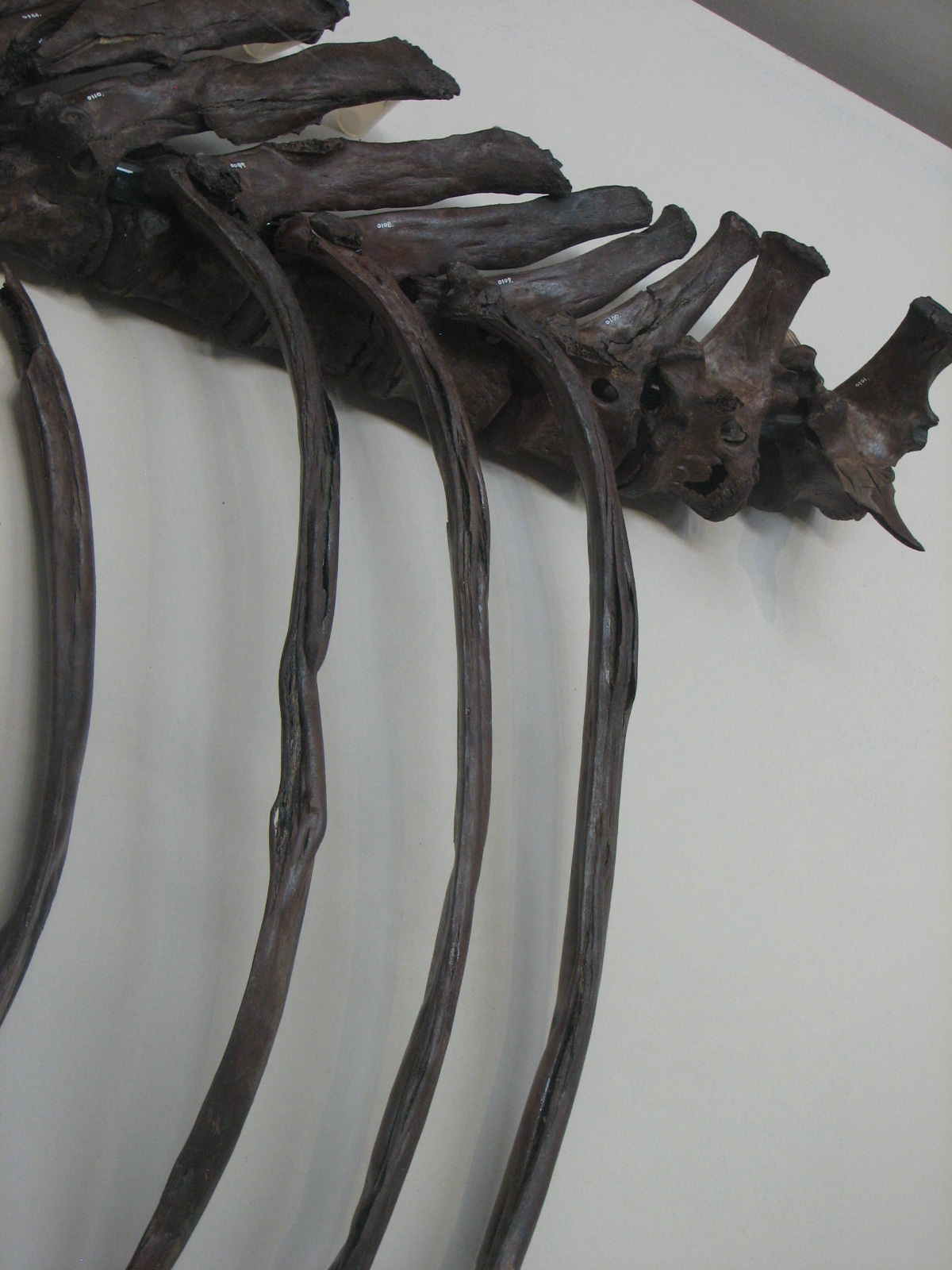
I invite anyone to school me on this phenomenon…

So how did it all get there?

There is little doubt the material in the well was artificially introduced. Nearly all the material is concentrated in the lower 2.5 m, with the very base of the deposit almost entirely ceramic vessels referred to as “earthquake spheres,” and the top of the deposits capped with stamped tile [4]. This suggests all the material was placed at the bottom of the well at one time [1], perhaps for “purification” [4]. There is the possibility that some (perhaps most) of the elements from the subterraneous critters (rodents, moles) may have been naturally introduced, but this can’t be tested without further data. Incidentally, for those concerned about dead animals at the bottom of a water source, it turns out that cold, stagnant water actually does a decent job of hindering decomposition. Besides, just about any natural body of water will have dead animals floating in it.

The inclusion of the child is also the subject of much speculation; one hypothesis suggests he or she may have been victim of drowning, subsequently offered as a sacrifice [1] [4].

Other hypotheses lack scientific rigor...

Hopefully, there will be more concrete explanations soon – allegedly, there is to be a professional publication on the excavation before the end of the year [3].


REFS

1. Gyeongju National Museum exhibition...

2. "Fell into a well Silla animals' exhibition

3. Gyeongju museum fell into a well Silla animals' exhibition

4. Gyeongju Museum exhibition "fell into a well Silla Animals' prepared

ANDREWS, P., 1995, Experiments in taphonomy: Journal of Archaeological Science, v. 22, p. 147–153.

ANDREWS, P., and COOK, J., 1985, Natural modifications to bones in a temperate setting: Man (New Series), v. 20, p. 675–691.

Saturday, March 26, 2011

Probably the Worst Healed Fracture I've Seen

Update: Thanks to reader Mark'o Plenty for pointing out that the femur discussed below is, in fact, clearly avian. And shame on me for blindly assuming it was mammal and never thinking otherwise. Fortunately, this narrows down the list of victims - for a femur that size, the only real likely candidate out here would be a Wild Turkey (Meleagris gallopavo). This would also greatly narrow the list of culprits responsible for the break - most likely, a motor vehicle is responsible, as the galliformes in this area have near-suicidal tendencies with respect to the highways (Scherzer and Peck, personal observation).

After subjecting this blog to what admittedly may qualify as criminal neglect, I return to the business of blogging, with a rather morbid “welcome back” post. I’ve been volunteering at local museum, and recently made an interesting recent discovery in the bone seen here:

That’s the femur of a small mammal – proximal end on the left, distal on the left, lateral side facing the camera - and it is abundantly clear that for a good portion of its life, that mammal was never having a good day. This femur experienced a complete fracture of the shaft – you can see a little window into the marrow cavity at the bottom – which significantly offset the bone, but the animal amazingly survived, and the bone healed. You can see the mass of regrowth where the bone healed along the shaft in the image above, and in more detail below. When examining the picture below, you should also make note of the femoral head (arrow) for later discussion:

The original length of the femur was 90 cm, and following the distortion, this was reduced to a little less than 75 cm, a reduction of almost 20% of the bone length. To put this in perspective, this would be like taking the thigh of the average American adult male (5’ 11”) and shortening it by roughly 3”. The image at left (from here) is probably a good comparison. You may also note some offsetting of the distal epiphysis, perhaps the result of crushing associated with the fracturing of the shaft. This damage to the epiphysis is seen in more detail in the image below:


So who was the victim?

One might naturally wonder what small mammal of Eastern Montana is stoic enough to put the Honey Badger to shame. Unfortunately, the damage to the femur seems to have inflicted some side effects that hinder proper identification. Based solely on gross osteology and original length, I was able to narrow down six possible taxa with the help of Wolniewicz’s field guide of mammal bones (2004): Virginia Opossum (Didelphis virginiana), Swift Fox (Vulpes velox)*, Domestic cat (Felis catus), Bobcat (Lynx rufus), Coyote (Canis latrans), and Domestic Dog (Canis lupus familiaris)**.

* Wolniewicz only provided images of Grey Fox (Urocyon cinereoargenteus) and Red Fox (Vulpes vulpes), but from my understanding, their ranges do not include Eastern Montana, where they are replaced by the Swift Fox, so I made an educated deduction.

** Neither did he include Domestic Dog or Grey Wolf (Canis lupus), but considering the resemblance of the femur to that of the Grey and Red Fox, I figured it was prudent to include the Domestic Dog as a possibility.

But then a visible problem arose - if you look at the femur dorsoventrally (at left), the mediolateral offsetting from the fracture seems relatively minor, hinting that the animal may have tried to use the limb following healing. But closer inspection of the proximal end suggests otherwise. The femoral head pointed out earlier (and visible in the top left of the image to the left), appears abnormally small, as can be seen by comparing it to the femoral head of some of the possible taxa:

(L to R) Domestic Cat, Bobcat, Coyote, Opossum. All to relative scale, all posterior view, femoral head at upper right. Ignore the lines. From Wolniewicz (2004).


In addition, all three trochanters (greater, lesser, and third) are practically absent. I noticed what appear to be rodent gnaw marks where the greater trochanter should be, but the lesser and third trochanters just plain aren’t there. This suggests the animal did not place weight on damaged limb after all, causing the associated muscles, and their respective trochanters, to atrophy as a result. This is good news for some of the taxa that were originally excluded due to their large size, namely the Bobcat and Coyote. A 90 cm femur is unlikely to come from an adult member of either of these taxa, and the fused epiphyses indicated this femur was not from a juvenile. However, a juvenile individual could have broken the femur, and the subsequent neglect would have left it matured but undersized (which is not uncommon). But this is bad news for me, since these trochanters are very useful in identifying taxa, so I was left with a short hand (no pun intended). One tantalizing clue that remains in an unusual groove in the lateral condyle (seen below). This groove is not diagnostic of any North American mammal I’m familiar with, and I didn’t see the feature in any of the images from Wolniewicz (2004). But I would love to hear I’m wrong***.

***Anyone? Anyone?


So what’s the taphonomic connection?

Well, none really. I considered weaving into a discussion on the taphonomic significance of broken bones, or the importance of pathologic features in the fossil record, but I didn’t want to distract from the main focus. A small mammal persevering in the harsh Eastern Montana climate with a deformed and vestigial hind limb is inspirational, if nothing else…


REFS

Wolniewicz, R. 2004. Field guide to skulls and bones of mammals of the northeastern United States, volume 2: the long bones. Richard Wolniewicz Publisher, Magnolia, MA, 97 p.